Anatomy Muscles Pelvis : Muscles of the Pelvic Floor - Anatomy & Function | Kenhub - The nerve supply to most of the muscles in this compartment is the obturator nerve, which arises from the lumbar plexus from.
Anatomy Muscles Pelvis : Muscles of the Pelvic Floor - Anatomy & Function | Kenhub - The nerve supply to most of the muscles in this compartment is the obturator nerve, which arises from the lumbar plexus from.. Key facts about the muscles of the pelvic floor. Learn about anatomy muscles pelvis with free interactive flashcards. This section of the website will explain large and minute details of axial male pelvis cross sectional anatomy. The floor of the pelvis is formed by the two muscles named levator ani and coccygeus. This mri pelvis cross sectional anatomy tool is absolutely free to use.
The muscles of the pelvis form its floor. A schematized 3d model of the pelvic diaphragm muscles (levator ani + coccygeus) and the obturator internus. • internal iliac (hypogastric) artery. The pelvic girdle consists of two symmetrical halves. Included within the chart are gorgeous illustrations of the pelvic diaphragm, sphincter muscles.
Hip Circle Progressions For Fun And Profit - Charlie Faraday from farm2.staticflickr.com Differences between the male pelvis and the female pelvis. Pelvic floor muscles located wholly within the pelvis. The muscles of the pelvis form its floor. Included within the chart are gorgeous illustrations of the pelvic diaphragm, sphincter muscles. Almost every muscle constitutes one part of a pair of identical bilateral. The muscles of the pelvis, hip and buttock anatomical chart shows how each muscle in this area of the body works with the others, and the various minor systems within the major ones. Anatomy ▶ pelvis ▶ muscles ▶ muscles of the pelvis. Define the pelvic girdle and describe the bones and ligaments of the pelvis.
Learn about anatomy muscles pelvis with free interactive flashcards.
Included within the chart are gorgeous illustrations of the pelvic diaphragm, sphincter muscles. Learn anatomy faster and remember everything you learn. The nerve supply to most of the muscles in this compartment is the obturator nerve, which arises from the lumbar plexus from. It originates from the pelvic outermost layer of the middle 3 sections of sacrum by 3 digitations. Key facts about the muscles of the pelvic floor. This anatomy section promotes the use of the terminologia anatomica, the international standard of anatomical nomenclature. Differences between the male pelvis and the female pelvis. The main functions of the neck muscles are to permit movements of the neck or head and to provide structural support of the head. There are many muscles that form the pelvic floor, including puborectalis, pubococcygeus, iliococcygeus and coccygeus. Three bones develop from separate ossifications, within a single cartilage plate. (1) the obturator internus and the piriformis, which are muscles of the lower extremity, and will be described with these (pages 476 and 477); The pelvic floor is also known as the pelvic diaphragm. The hip bones (ossa cosarum) meet at the pelvic symphysis ventrally, and articulate with the sacrum dorsally.
This mri pelvis cross sectional anatomy tool is absolutely free to use. It originates from the pelvic outermost layer of the middle 3 sections of sacrum by 3 digitations. .anatomy of the pelvicgirdle better manage your patients with pelvicgirdle dysfunction by knowing the interaction of the bones, joints, ligaments, and muscles around the pelvis powered by physiopedia start course presented by: Explain the three regions of the hip bone and identify their bony landmarks. Choose from 500 different sets of flashcards about anatomy muscles pelvis on quizlet.
MRI pelvis anatomy | free male pelvis axial anatomy from mrimaster.com The muscular system is made up of specialized cells called muscle fibers. The floor of the pelvis is formed by the two muscles named levator ani and coccygeus. (1) the obturator internus and the piriformis, which are muscles of the lower extremity, and will be described with these (pages 476 and 477); This mri pelvis cross sectional anatomy tool is absolutely free to use. These muscles all serve as adductors of the thigh, but also serve as important stabilizers of the pelvis and work to maintain balance of the pelvis on the lower limb during gait. ƒ important to understand normal anatomy. Deborah riczo dr deborah riczo is a specialist in pelvic •. .anatomy of the pelvicgirdle better manage your patients with pelvicgirdle dysfunction by knowing the interaction of the bones, joints, ligaments, and muscles around the pelvis powered by physiopedia start course presented by:
ƒ pelvic floor dysfunction is common and.
This article reviews the anatomical and functional information of the gastrocnemius muscle, its embryological derivation. Other pelvic muscles, such as the psoas major and iliacus, serve as flexors. The muscles within the pelvis may be divided into two groups: • internal iliac (hypogastric) artery. The main functions of the neck muscles are to permit movements of the neck or head and to provide structural support of the head. These muscles origin in continuity from the body of the pubis, along a tendinous arch over the obturator internus fascia, and the ischial spine. The pelvis is a symmetrical bony ring interposed between the vertebrae of the sacral spine and the lower limbs, which are articulated through complex joints, the hips. Key facts about the muscles of the pelvic floor. Cross the ls joint onto the trunk 2. The pelvis and the pelvic floor muscles seal the abdominal and pelvic cavity in a caudal direction; Muscles, connected to bones or internal organs and blood vessels, are in charge for movement. There are around 650 skeletal muscles within the typical human body. Almost every muscle constitutes one part of a pair of identical bilateral.
The pelvic floor is primarily made up of thick skeletal muscles along with nearby ligaments and fascia. The nerve supply to most of the muscles in this compartment is the obturator nerve, which arises from the lumbar plexus from. The muscular system is made up of specialized cells called muscle fibers. The floor of the pelvis is formed by the two muscles named levator ani and coccygeus. The muscles of the pelvis form its floor.
Structure and Function of the Hip | Musculoskeletal Key from musculoskeletalkey.com Learn anatomy faster and remember everything you learn. It affects the entire lower limb and the movement of the hip and the lumbar area. ƒ important to understand normal anatomy. A schematized 3d model of the pelvic diaphragm muscles (levator ani + coccygeus) and the obturator internus. This is a table of skeletal muscles of the human anatomy. Muscles, connected to bones or internal organs and blood vessels, are in charge for movement. Almost every movement in the body is the outcome of muscle contraction. Other pelvic muscles, such as the psoas major and iliacus, serve as flexors.
The pelvis and the pelvic floor muscles seal the abdominal and pelvic cavity in a caudal direction;
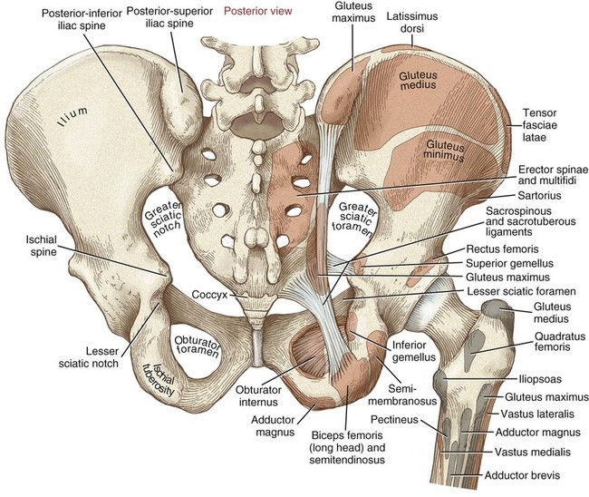
The muscles within the pelvis may be divided into two groups: Deborah riczo dr deborah riczo is a specialist in pelvic •. Muscles, connected to bones or internal organs and blood vessels, are in charge for movement. These muscles, including the gluteus maximus and the hamstrings, extend the thigh at the hip in support of the body's weight and propulsion. The pelvic girdle consists of two symmetrical halves. The muscles of the pelvis, hip and buttock anatomical chart shows how each muscle in this area of the body works with the others, and the various minor systems within the major ones. The sacrospinous and sacrotuberous ligaments also help to define two openings on the posterolateral sides of the pelvis through which muscles. It attaches to the walls of the lesser pelvis, separating the in this article, we shall look at the anatomy of the muscles that make up the inferior lining of the cavity; The pelvic floor is primarily made up of thick skeletal muscles along with nearby ligaments and fascia. And pathophysiology to properly care for women with these conditions. These muscles origin in continuity from the body of the pubis, along a tendinous arch over the obturator internus fascia, and the ischial spine. The main functions of the neck muscles are to permit movements of the neck or head and to provide structural support of the head. Key facts about the muscles of the pelvic floor.


0 Komentar